
More Information
Submitted: October 20, 2026 | Accepted: October 27, 2026 | Published: October 29, 2026
Citation: Cojocaru AV, Balint S. The Dynamics of a Continuous-time Artificial Hopfield Neural Network after a Rosehip Neuron Inhibits a Pyramidal Neuron, using a Sigmoid Input-output Function. Int J Phys Res Appl. 2026; 9(10):331-340. Available from:
https://dx.doi.org/10.29328/journal.ijpra.1001168
DOI: 10.29328/journal.ijpra.1001168
Copyright license: © 2026 Cojocaru AV, et al. This is an open access article distributed under the Creative Commons Attribution License, which permits unrestricted use, distribution, and reproduction in any medium, provided the original work is properly cited.
Nomenclature: Continuous-time Hopfield neural network; Rosehip neuron; Pyramidal neuron; Sigmoid activation function; Neural inhibition; Equilibrium stability

The Dynamics of a Continuous-time Artificial Hopfield Neural Network after a Rosehip Neuron Inhibits a Pyramidal Neuron, using a Sigmoid Input-output Function
Andreea V Cojocaru* and Stefan Balint*
Department of Computer Science, West University of Timisoara, Blvd. V. Parvan 4, 300223 Timisoara, Romania
*Address for Correspondence: Andreea V. Cojocaru, Department of Computer Science, West University of Timisoara, Blvd. V. Parvan 4, 300223 Timisoara, Romania, Email: [email protected]
Stefan Balint, Department of Computer Science, West University of Timisoara, Blvd. V. Parvan 4, 300223 Timisoara, Romania, Email: [email protected]
Using a system of a two-dimensional Hopfield continuous-time artificial network with self-connection and without constant external electrical input, in this paper the computed voltage dynamics are presented after the inhibition of a pyramidal neuron by a rosehip neuron. Because the rest state of the pyramidal neuron is not conserved after the inhibition, a way to find the transfer matrix elements that can be used after the inhibition is given, which ensures that the pyramidal neuron rest state is conserved after the inhibition.
In the real world, there are biological neural networks (nervous system) and artificial (human-made) neural networks. For the biological ones, the mathematical framework was developed by Hodgkin and Huxley (Nobel Prize in 1963) [1], and for the artificial ones, it was developed by Hopfield (Nobel Prize in 2024) [2]. The aim of our research is to find how the behavior of a neural network composed of a rosehip and a pyramidal biological cell can be mirrored in a Hopfield-type artificial neural network. This aim is not just an academic curiosity, considering the fact that measurements in the case of rosehip and pyramidal biological cells are made using artificial neural networks. Rosehip neurons are inhibitory GABAergic neurons, present in the first layer (the molecular layer) of the human cerebral cortex. They make up about 10-15% of all inhibitory neurons in Layer 1 [3]. Neurons of this type (having “large ‘rosehip’-like axonal buttons and compact arborization”) exist in humans, but have not been reported in rodents [2]. Rosehip neurons are named after the rosehip fruit due to the axon terminal’s resemblance to its berries. These rosehip cells show an immunohistochemical profile (GAD 1 +CCK +, CNR1-SST-CALB2- PVALB-) matching a single transcriptomically defined cell type whose specific molecular marker signature is not seen in mouse cortex. Rosehip cells in layer 1 make homotypic gap junctions, predominantly target apical dendritic shafts of layer 3 pyramidal neurons, and inhibit back-propagating pyramidal action potentials in micro domains of the dendritic tuft. These cells are therefore positioned for potent local control of distal dendritic computation in cortical pyramidal neurons. An international group of scientists discovered Rosehip neurons and announced their discovery in August 2018. These authors contributed equally to this work: Eszter Boldog (University of Szeged, Szeged, Hungary), Trygve E. Bakken (Allen Institute for Brain Science, Seattle, WA, United States), and Rebecca D. Hodge (Allen Institute for Brain Science, Seattle, WA, United States). They identified this cell with the help of RNA sequencing [4]. Continuous-time artificial Hopfield neural networks claims to be mathematical descriptions of voltage propagation appearing in artificial neural networks. If this description is also valid for the biological nervous system, then the voltage propagation in a rosehip-pyramidal nervous cell network has to be mirrored with accuracy in this framework. In particular, it has to reflect that rosehip nervous cell can inhibit a pyramidal cell and can describe the voltage dynamics also after inhibition. The inhibition problem was solved in [7,8]. The aim of this paper is, to analyze the voltage dynamic after the inhibition and identify a way to find the transfer matrix elements which has to be used after the inhibition moment which assure that the pyramidal neuron rest state is conserved.
In this paper, it is assumed that the system of differential equations governing the voltage dynamics of the rosehip-pyramidal neuron is a two dimensional Hopfield continuous-time artificial network with self-connection and without constant external electrical input, which is
(1.1)
This is due to the anomalies concerning the necessary external current magnitude, revealed in [5,6].
In (1.1): x[V] is the voltage of the ‘pyramidal’ neuron (neuron whose voltage has to be shifted), y[V] is the voltage of the ‘rosehip’ neuron. In this computation, the numerical values of the time constant are: a1 = 100 [s]-1 (pyramidal cell), a2 = 136 [s]-1 (rosehip cell). The function is the input-output function. An input-output function is a mathematical “gate” between the input feeding and its output going. The input-output activation functions are used to calculate some intermediate functions in the hidden layer, which are then used to calculate an output. According to [7,8] the numerical values of transfer matrix elements T11, T22 are found using formulas: and with
In [7], in the case of the sigmoid input-output function, the transfer coefficient T21 was taken as T21 = 4.3.
In [8], for 9 input/output function, the following elements of the transfer matrix can be found:
1. Sigmoid input-output function:
threshold voltage inhibition: T11 =- 15.58413113, T12 =-43, T22 =- 19.73027788, T21 = 4.3
spike voltage inhibition;
hyperpol. voltage inhib.;
2. Sigmoid*(1-Sigmoid) input-output function:
threshold voltage inhibition;
spike voltage inhibition ;
hyperpol. voltage inhib.;
3. Tanh. input-output function:
threshold voltage inhibition;
spike voltage inhibition ;
hyperpol. voltage inhib.;
4. Tanh-Derivative input-output function:
threshold voltage inhibition;
spike voltage inhibition ;
hyperpol. voltage inhib.;
5. Linear input-output function:
threshold voltage inhibition; T11 = 100, T12 = 365, T22 = 136, T21 = 1
spike voltage inhibition ; T11 = 100, T12 = 1547, T22 = 136, T21 = 1
hyperpol. voltage inhib.; T11 = 100, T12 = -20, T22 = 136, T21 = 1
6. Leaky-ReLU input-output function:
threshold voltage inhibition; T11 = 1000, T12 = -560 , T22 = 1360, T21 = -825
spike voltage inhibition ; T11 = 1000, T12 = 634.5, T22 = 1360, T21 = -825
hyperpol. voltage inhib.; T11 = 1000, T12 = -1229, T22 = 1360, T21 = -825
7. Swish input-output function:
threshold voltage inhibition;
spike voltage inhibition ;
hyperpol. voltage inhib.;
8. ELU input-output function:
threshold voltage inhibition;
spike voltage inhibition;
hyperpol. voltage inhib.;
9. GELU input-output function:
threshold voltage inhibition;
spike voltage inhibition;
hyperpol. voltage inhib.;
The above-presented transfer matrix coefficients are not unique. There exist other choices of the coefficient T21 that lead to different values of T12 for a given input- output function. According to our computation concerning the voltage dynamics, what is certain is that for the above-presented transfer matrix coefficients, the inhibition of the pyramidal neuron takes place in 0.001 [s].
In the present paper, we analyze the voltage dynamics after 0.001[s] in the case of a sigmoid input-output function. Remember that the sigmoid input-output function is and its graph is represented in the next figure.
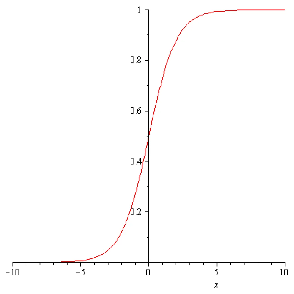
Figure 1.1: The graph of the sigmoid function .
2. The voltage dynamics after the inhibition of the threshold voltage of a pyramidal neuron in case of the sigmoid input-output function
In case of the threshold voltage inhibition the voltage dynamics during the first second are represented in the next figure.
This figure shows that, after the inhibition of the threshold voltage of the pyramidal neuron, which takes place at 0.001[s], the voltage of the pyramidal neuron decreases towards the value of -0.262095713 [V], which is much less than its rest voltage. Moreover, the voltage of the rosehip neuron decreases towards the value of =-0.0567318469 [V].
The explanation is that the voltage state x = -0.262095713 [V], y = -0.05673184699[V] is a steady state of the system (1.1). The eigenvalues of the Jacobi matrix in this steady state are . Therefore, the steady state x = -0.262095713 [V], y = -0.05673184699 [V] is a stable node (sink); see [5,6]. The voltage field around this steady state is represented on the next figure.
It follows that, if the elements of the transfer matrix are maintained constants after the moment of inhibition (0.001[s]) the computed dynamic of the pyramidal neuron voltage, after the inhibition of threshold potential is the consequence of a pure mathematic phenomena, and the rest voltage of pyramidal neuron is not conserved.
A way to maintain the voltage of the pyramidal neuron constant is to shift the values of the transfer matrix after the accomplishment of the inhibition. This can be made for example, by taking in system (1.1), T12 = T21 = 0 and taking the initial conditions t = 0.001[s], x(0.001) = -0.0754472959796389231[V], y(0.001) = -0.0550321350788334010[V]. The computed voltage dynamics in this case are presented in the next figure:
This figure shows that, after the switch of the transfer matrix element value, at the moment of time 0.001[s], the pyramidal and the rosehip neuron voltages evolve towards the corresponding rest voltages, -0.075[V] and -0.07[V], respectively. The explanation is that if T12 = 0 and T21 = 0, then the voltage state x = -0.075 [V], y = -0.07 [V] is a steady state of the system (1.1)The eigenvalues of the Jacobian matrix in the steady state are , . Therefore, the rest state x = -0.075 [V], y = -0.07 [V] is a stable node (a sink) in the sense of [5,6] and the voltage field around the rest state is that represented in the next figure:
It follows that, if the elements of the transfer matrix are switched at the moment of inhibition (i.e., 0.001[s]), the pyramidal and the rosehip neuron voltages evolve towards the corresponding rest voltages, -0.075[V] and -0.07[V], respectively.
In a Hopfield-type artificial neural network, the coefficients of the transfer matrix (more commonly referred to as the weight matrix or interconnection matrix), is usual do not have static numerical values. Instead, their values are computed dynamically based on the specific memory patterns you want the network to store.
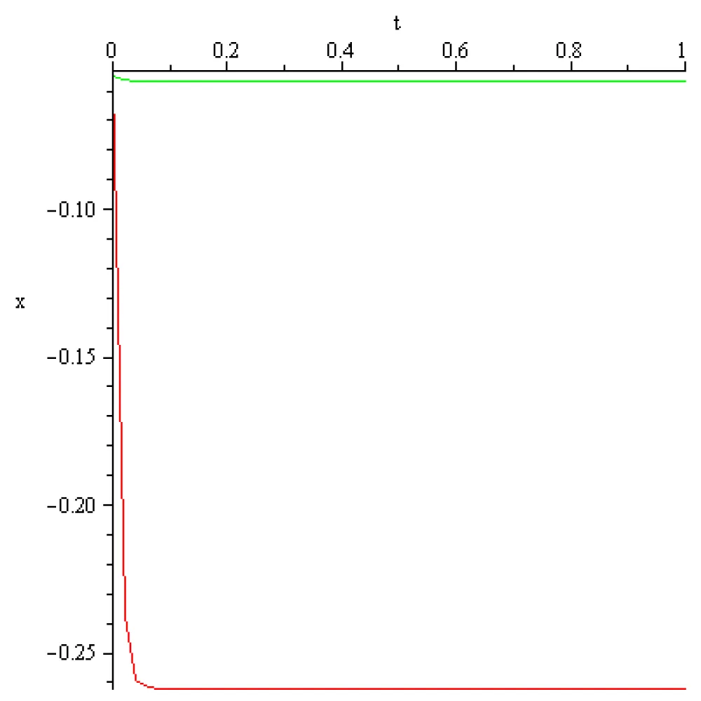
Figure 2.1: The pyramidal neuron (red) and the rosehip neuron(green) dynamic in case T12 = -43, T21 = 4.3, t = 1[s], x(0) = -0.055, [V], y(0) = -0.055[V], x(1) = -0.262095713 [V], y(1) = -0.0567318469[V].
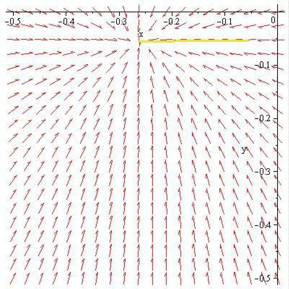
Figure 2.2: The voltage field around the steady state x = -0.262095713 [V], y = -0.05673184699[V].
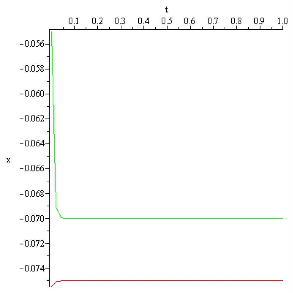
Figure 2.3: The pyramidal neuron (red) and the rosehip neuron(green) voltage dynamic after the shift T12 = 0, T21= 0, t = 0.001…1[s], x(0.001) = -0.0754472959796389231[V], y(0.001) = -0.0550321350788334010[V], x(1) = -0.075 [V], y(1) = -0.07 [V].
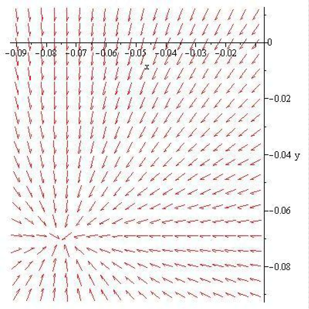
Figure 2.4: The voltage field around the rest state x = -0.075 [V], y = -0.07 [V].
3. The voltage dynamics after the inhibition of the spike voltage of a pyramidal neuron in case of the sigmoid input-output function
In the case of spike voltage inhibition and the voltage dynamics during the first second are represented in the next figure.
This computation shows that, after the inhibition of the spike voltage of the pyramidal neuron, which takes place at 0.001[s], the voltage of the pyramidal neuron decreases towards the value -0.8513651515 [V], which is much less than its rest voltage, and the voltage of the rosehip neuron decreases towards the value of -0.06087238335 [V].
The explanation is that the voltage state x=-0.8513651515 [V], y =-0.06087238335 [V] is a steady state of the system (1.1). The eigenvalues of the Jacobi matrix in this steady state are , Therefore, according to [5,6], the steady state x = -0.8513651515 [V], y = -0.06087238335 [V] is a stable node (sink), and the voltage field around this steady state is that represented in the next figure.
It follows that the computed dynamics of the pyramidal neuron voltage, after the inhibition of the spike potential, is a mirror of a pure mathematical fact and is different from the rest voltage of the pyramidal neuron.
A way to maintain the rest voltage of the pyramidal neuron after the spike voltage inhibition is to shift the values of the transfer matrix after the accomplishment of the inhibition. This can be done, for example, by taking in system (1.1), T12 = T21 = 0 and taking the initial conditions t = 0.001[s], x(0.001) = -0.0750545288420509660[V], y(0.001)=-0.0550006881737295230[V]. The computed voltage dynamics after the shift are presented in the next figure:
This computation shows that, after the switch of the transfer matrix element value, at the moment of time 0.001[s], the pyramidal and the rosehip neuron voltages evolve towards the corresponding rest voltages, -0.075[V] and -0.07[V], respectively. The explanation is that if T12 = 0 and T21 = 0, then the voltage state x = -0.075 [V], y = -0.07 [V] is a steady state of the system (1.1). The eigenvalues of the Jacobian matrix in the steady state are , . Therefore, the rest state x = -0.075 [V], y = -0.07 [V] is a stable node (sink) in the sense of [5,6]. The voltage field around this steady state is represented in the next figure:
It follows that, if the elements of the transfer matrix are switched at the moment of inhibition (i.e., 0.001[s]), the pyramidal and the rosehip neuron voltage evolve towards the corresponding rest voltages, -0.075[V] and -0.07[V], respectively.
In a Hopfield-type artificial neural network, the coefficients of the transfer matrix (more commonly referred to as the weight matrix or interconnection matrix), is usual do not have static numerical values. Instead, their values are computed dynamically based on the specific memory patterns you want the network to store.
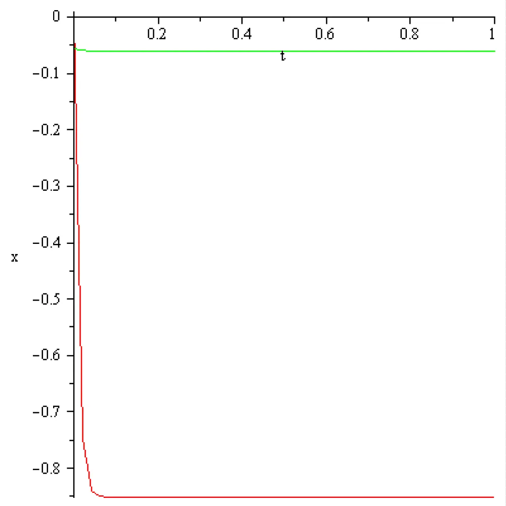
Figure 3.1: The pyramidal neuron (red) and the rosehip neuron(green) dynamic in case T12 = -166, T21= 4.3, t = 1[s], x(0) = 0.01 [V], y(0) = -0.055 [V], x(1) = -0.8513651515 [V], (1) = -0.06087238335 [V].
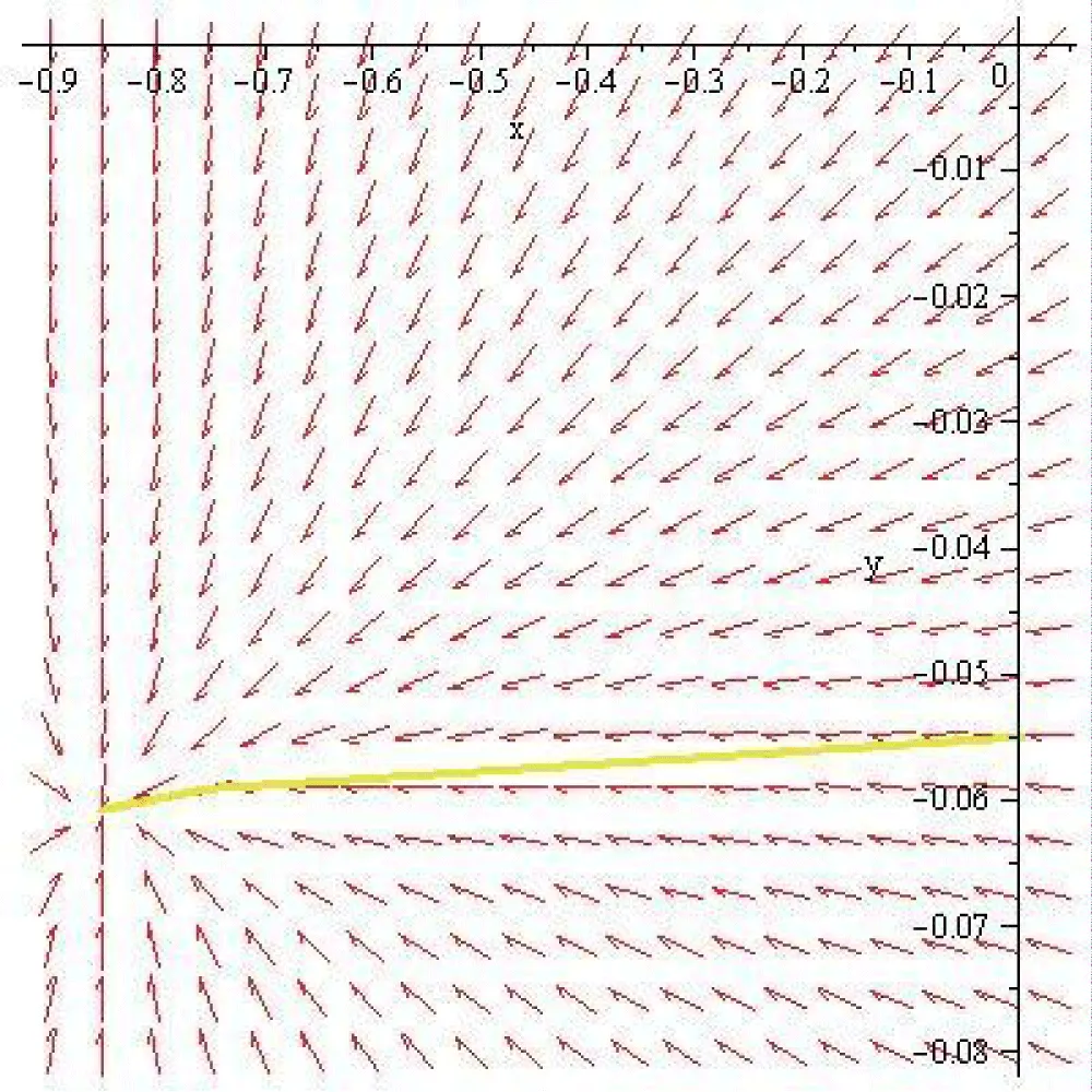
Figure 3.2: The voltage field around the steady state x = -0.8513651515[V], y = -0.06087238335[V].
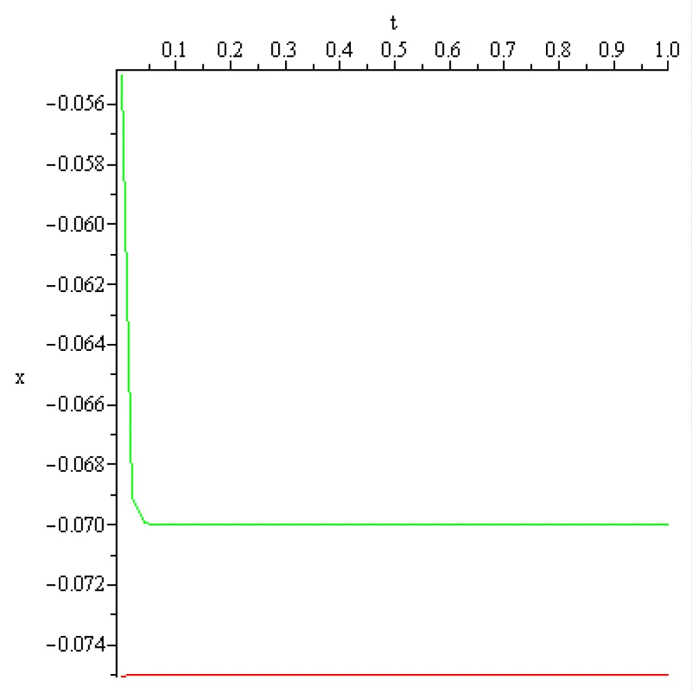
Figure 3.3: The pyramidal neuron (red) and the rosehip neuron(green) dynamic in case T12= 0, T21 = 0, t = 1[s], x(0.001) = -0.0750545288420509660[V], y(0.001) = -0.0550006881737295230[V].
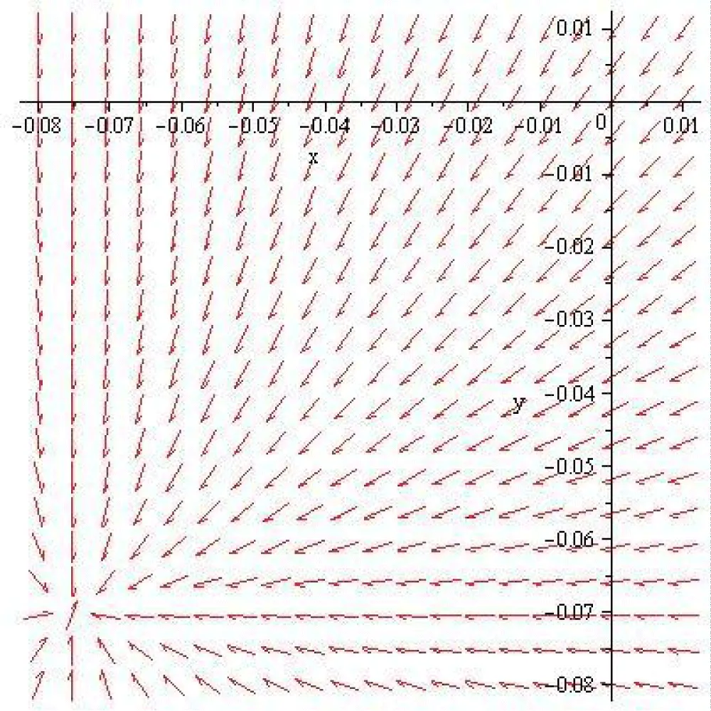
Figure 3.4: The voltage field around the rest state x = -0.075 [V], y = -0.07 [V].
4. The voltage dynamics after the inhibition of the hyperpolarization voltage of a pyramidal neuron in case of the sigmoid input-output function
In the case of hyperpolarization voltage inhibition , and the voltage dynamics during the first second are represented in the next figure.
This computation shows that, after the inhibition of the hyperpolarization voltage of the pyramidal neuron, which takes place at 0.001[s], the voltage of the pyramidal neuron increases towards the value -0.05674815844 [V], which is much more than its rest voltage, and the voltage of the rosehip neuron evolves towards the value of -0.05517671254[V]. The explanation is that the voltage state x = -0.05674815844 [V], y = -0.05517671254 [V] is a steady state of the system (1.1.) The eigenvalues of the Jacobi matrix in the steady state are . Therefore, according to [5,6], the steady state x = -0.05674815844 [V], y = -0.05517671254 [V] is a stable node (sink), and the voltage field around this steady state is that represented in the next figure.
It follows that the computed dynamics of the pyramidal neuron voltage, after the inhibition of the hyperpolarization potential, is a mirror of a pure mathematical fact and is different from the rest voltage of the pyramidal neuron.
A way to maintain the rest voltage of the pyramidal neuron constant after the spike voltage inhibition is to shift the values of the transfer matrix after the accomplishment of the inhibition. This can be done, for example, by taking in system (1.1), T12 = T21 = 0 and taking the initial conditions t = 0.001, x(0.001)=-0.0750545288420509660[V], y(0.001)=-0.0550006881737295230[V]. The computed voltage dynamics after the shift are presented in the next figure:
This figure shows that, after the switch of the transfer matrix element value, at the moment of time 0.001[s], the pyramidal and the rosehip neuron voltages evolve towards the corresponding rest voltages, -0.075[V] and -0.07[V], respectively. The explanation is that if T12 = 0 and T21 = 0,then the voltage state x = -0.075 [V], y = -0.07 [V] is a steady state of the system (1.1). The eigenvalues of the Jacobian matrix in the steady state are . Therefore, the steady state x = -0.075 [V], y = -0.07 [V] is a stable node (sink) in the sense of [5,6]. The voltage field around this steady state is represented in the next figure:
It follows that, if the elements of the transfer matrix are switched at the moment of inhibition (i.e., 0.001[s]) the pyramidal and the rosehip neuron voltage evolve towards the corresponding rest voltages, -0.075[V] and -0.07[V], respectively.
In a Hopfield-type artificial neural network, the coefficients of the transfer matrix (more commonly referred to as the weight matrix or interconnection matrix), is usual do not have static numerical values. Instead, their values are computed dynamically based on the specific memory patterns you want the network to store.
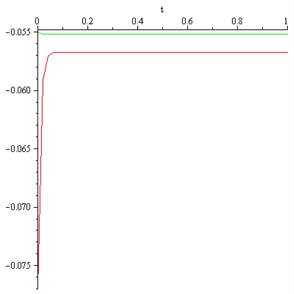
Figure 4.1: The pyramidal neuron (red) and the rosehip neuron(green) dynamic in case T12= 0, T21= 0, t = 1[s], x(0.001) = -0.0750545288420509660[V], y(0.001) = -0.0550006881737295230[V], x(1) = -0.75, y(1) = -0.07.
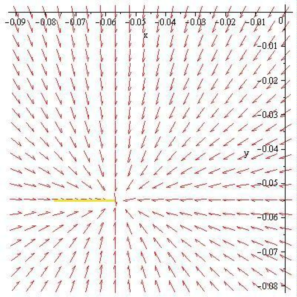
Figure 4.2: The voltage field around the rest state x = -0.075 [V], y = -0.07 [V].
Computation shows that, in the case of a sigmoid input-output function, after the inhibition of the pyramidal neuron voltage, which takes place at 0.001[s], maintaining the elements of the transfer matrix constants the voltage of the pyramidal neuron evolves toward the steady state of the system.
By changing at 0.001[s], in an appropriate way (T12 = 0, T21 = 0), the elements of the transfer matrix, the pyramidal neuron voltage evolves toward the rest state.
a). The use of an artificial Hopfield neural network with self-connection is legitimated by the fact that the rest state xo [V] and the time constant is different from zero in case of rosehip and pyramidal nervous cells.
b). It turns out that the prior giving of the elements T21 and T12 of the transfer matrix and the exclusive use of the constant external impulse current input in [5] and [6] leads to a necessary current of magnitude that is higher than the magnitude reported in the literature.
c). The presented computed transfer matrix coefficients are not unique. There exist other choices of coefficient T21 which lead to different values of T12 for the sigmoid input- output function. This is also true in the case of switching the values of these coefficients.
d). Computation shows that the shift of coefficients T21 and T12 is necessary at the moment of inhibition. Otherwise, the voltage dynamics do not evolve towards the rest state voltage, maintained by ion pumps and the selective permeability of the membrane. For this ionic mechanisms discovery, the Nobel Prize in Physiology or Medicine 1963 was awarded jointly to Sir John Carew Eccles, Alan Lloyd Hodgkin, and Andrew Fielding Huxley [1].
Authors’ contribution: The authors contributed equally to the realization of this work. All authors have read and agreed to the published version of the manuscript.
Funding: This research did not receive any specific grant from funding agencies in the public, commercial or not-for-profit sectors.
Data availability statement: The original contributions presented in the study are included in the article; further inquiries can be directed to the corresponding author.
Conflicts of interest: The authors declare no conflicts of interest.
- McRae M. Scientists have found a new type of brain cell and it looks like it's unique to humans. ScienceAlert [Internet]. 2018 [cited 2018 Sep 1]. Available from: https://www.sciencealert.com/rosehip-neuron-discovery-absent-in-mouse-models
- Boldog E, Bakken TE, Hodge RD, Novotny M, Aevermann BD, Baka J, et al. Transcriptomic and morphophysiological evidence for a specialized human cortical GABAergic cell type. Nat Neurosci. 2018 Sep;21(9):1185-95. Available from: https://doi.org/10.1038/s41593-018-0205-2
- Cojocaru AV, Balint S. Mirroring the pyramidal-neuron cell behavior in the continuous-time artificial Hopfield neural network: What problems arise? Int J Phys Res Appl. 2026;9(6):190-201. Available from: https://doi.org/10.29328/journal.ijpra.1001156
- Cojocaru AV, Balint S. Mirroring the rosehip-neuron cell behavior in the continuous-time artificial Hopfield neural network: What problems arise? Int J Phys Res Appl. 2026;9(6):162-89. Available from: https://doi.org/10.29328/journal.ijpra.1001155
- Cojocaru AV, Balint S. Mirroring the inhibition of a pyramidal neuron by a rosehip neuron in continuous-time artificial Hopfield neural network [preprint]. ResearchGate. 2026 Sep. Available from: https://doi.org/10.13140/RG.2.2.24383.06569